Best Physeal Bar Treatment in Indore (ग्रोथ प्लेट पर हड्डी का पुल)
Physeal Bar & Growth Plate Arrest in Children
फ्रैक्चर जुड़ गया, फिर भी पैर टेढ़ा हो रहा है? | ग्रोथ प्लेट पर बना हड्डी का पुल इसकी वजह हो सकता है | Indore
The fracture healed — so why is the leg bending? | A bar of bone across the growth plate is often the answer | Indore
Physeal Bar क्या है?
What is a Physeal Bar?
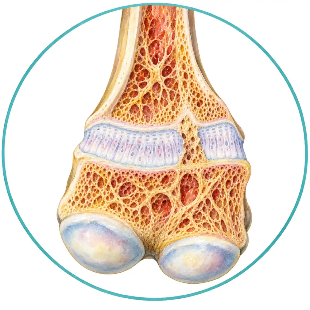
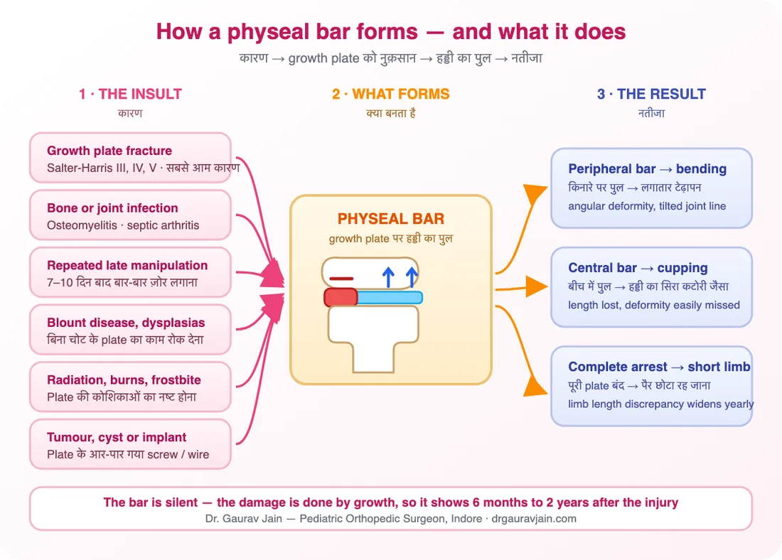
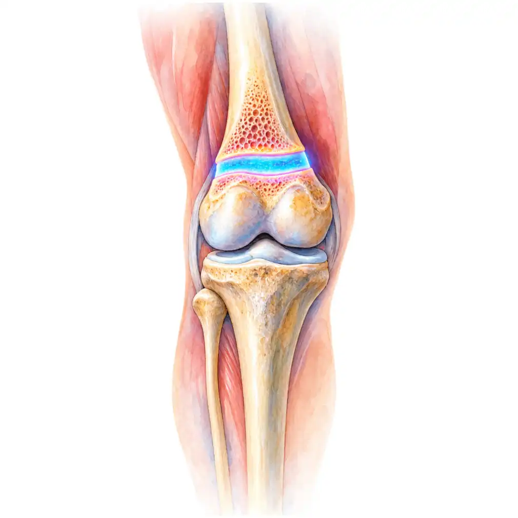
बच्चों की हड्डी सिरों के पास मौजूद एक नरम cartilage की पट्टी से लंबी होती है, जिसे growth plate (physis) कहते हैं। चोट, संक्रमण या किसी और नुक़सान के बाद इस पट्टी के किसी हिस्से में cartilage की जगह सख़्त हड्डी का पुल बन जाता है — इसी को physeal bar कहते हैं।
A child's bone grows in length from a band of soft cartilage near each end called the growth plate, or physis. After an injury, an infection or some other insult, part of that band can be replaced by a bridge of solid bone — and that bridge is a physeal bar.
🎯 असली दिक़्क़त यह है🎯 Why that one bridge matters so much
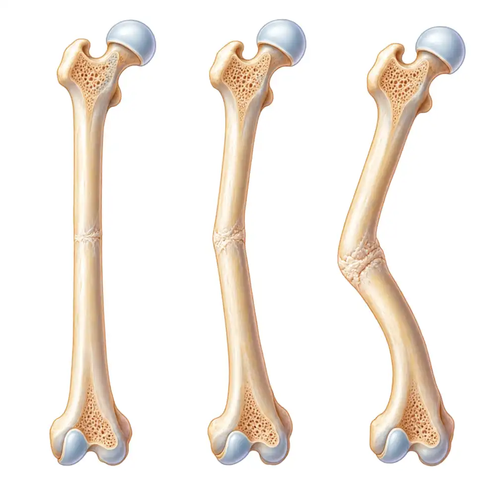
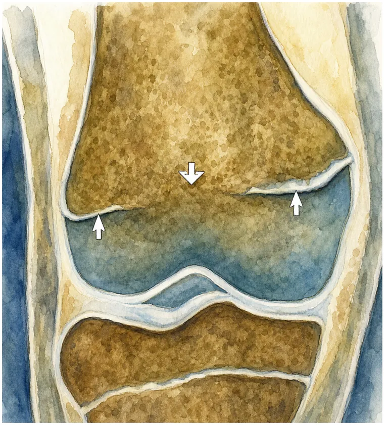
जहाँ bar बन गया, वहाँ हड्डी बढ़ना बंद कर देती है — लेकिन growth plate का बाक़ी हिस्सा बढ़ता रहता है। नतीजा यह होता है कि हड्डी एक तरफ़ से खिंची रहती है और दूसरी तरफ़ से बढ़ती है, इसलिए वह टेढ़ी होती चली जाती है। अगर पूरी plate पर bar बन जाए तो वह हड्डी बढ़ना ही बंद कर देती है और पैर छोटा रह जाता है।Wherever the bar sits, growth stops — but the rest of the plate carries on. The bone is therefore tethered on one side and free on the other, so it bends a little more with every centimetre the child grows. If the bar covers the whole plate, that bone simply stops lengthening and the limb is left short.
🕒 यह देर से क्यों दिखता है🕒 Why it shows up so late
Bar बनते ही कुछ नहीं दिखता, क्योंकि बिगाड़ चोट से नहीं — growth से पैदा होता है। इसलिए फ्रैक्चर ठीक होने के 6 महीने से 2 साल बाद जाकर पैर टेढ़ा दिखना शुरू होता है, तब तक ज़्यादातर परिवार चोट को भूल चुके होते हैं। यही कारण है कि growth plate की चोट के बाद follow-up इतना ज़रूरी है।Nothing happens the day the bar forms, because the damage is not done by the injury — it is done by growth. The bending usually becomes visible six months to two years after the fracture healed, by which time most families have stopped thinking about it. That single fact is why follow-up after a growth plate injury matters more than almost anything else on this page.
Bar कहाँ बना है — इसी से तय होता है क्या बिगड़ेगा
Where the Bar Sits Decides What Goes Wrong
एक ही बीमारी, पर तीन बिल्कुल अलग नतीजे — और यह पूरी तरह इस बात पर निर्भर करता है कि हड्डी का पुल growth plate के किस हिस्से में बना है।
One condition, three quite different outcomes — and which one a child gets depends entirely on where in the plate the bridge of bone happens to sit.
Peripheral bar — किनारे परPeripheral bar - at the edge
Plate के किनारे बना पुल। एक तरफ़ बढ़त रुकती है, दूसरी चलती रहती है — इसलिए हड्डी लगातार टेढ़ी (angular deformity) होती जाती है। घुटने के पास हो तो पैर बाहर या अंदर मुड़ने लगता है।A bridge at the rim of the plate. One side stops, the other keeps going, so the bone bends further with every month of growth — a progressive angular deformity. Near the knee it shows as a leg drifting into bow or knock-knee.
Central bar — बीच मेंCentral bar - in the middle
Plate के बीचोंबीच पुल। किनारे बढ़ते रहते हैं और बीच रुका रहता है, जिससे हड्डी का सिरा कटोरी जैसा (cupping/tenting) हो जाता है। बाहर से टेढ़ापन कम दिखता है, पर लंबाई रुक जाती है।A bridge in the middle of the plate. The rim keeps growing around a stalled centre, so the bone end becomes cupped or tented. There is less visible bending, but length is lost — and it is easily missed for that reason.
Complete arrest — पूरी plateComplete arrest - the whole plate
पूरी growth plate बंद। हड्डी टेढ़ी नहीं होती, पर बढ़ना ही रुक जाता है — और दूसरा पैर बढ़ता रहता है, इसलिए पैरों की लंबाई का अंतर (limb length discrepancy) हर साल बढ़ता जाता है।The whole plate closes. The bone does not bend, it simply stops — while the other limb carries on growing, so the difference in leg length widens every year until skeletal maturity.
⚠️ जितनी उम्र बची है, नुक़सान उतना बड़ा⚠️ The younger the child, the bigger the eventual problem
4 साल के बच्चे में बना bar 14 साल के बच्चे के मुक़ाबले कहीं ज़्यादा नुक़सान करता है, क्योंकि उसके पास बढ़ने के लिए बहुत साल बचे हैं। इसीलिए छोटे बच्चों में इलाज जल्दी और ज़्यादा गंभीरता से किया जाता है, भले ही आज विकृति मामूली दिख रही हो।A bar in a four-year-old does far more harm than the same bar in a fourteen-year-old, simply because there are so many growing years left for it to work in. That is why small children are treated earlier and more aggressively, even when today's deformity looks minor.
Physeal Bar क्यों बनता है
Why a Physeal Bar Forms
Growth plate की चोट (सबसे आम)A growth plate fracture - by far the commonest
Plate के आर-पार जाने वाला फ्रैक्चर ठीक होते समय दोनों तरफ़ की हड्डी आपस में जुड़ जाती है और वहीं पुल बन जाता है। घुटने के ऊपर (distal femur) और टखने के पास (distal tibia) की चोटें सबसे ज़्यादा ख़तरनाक हैं।A fracture that runs across the plate lets bone on either side unite through it as it heals, and the bridge forms there. Injuries just above the knee and around the ankle are the most dangerous of all.
हड्डी या जोड़ का संक्रमणBone or joint infection
शिशु अवस्था में osteomyelitis या septic arthritis growth plate का हिस्सा नष्ट कर सकता है। इन बच्चों में विकृति सबसे गंभीर होती है, क्योंकि बढ़ने के लिए पूरी उम्र बाक़ी होती है।Osteomyelitis or septic arthritis in infancy can destroy part of the plate outright. These children end up with the worst deformities, because a whole childhood of growth is still ahead of them.
बार-बार या देर से की गई manipulationRepeated or late manipulation
Growth plate की चोट को 7–10 दिन बाद दोबारा-दोबारा ज़ोर लगाकर सीधा करने की कोशिश plate को और नुक़सान पहुँचाती है। यह उन कुछ कारणों में है जिन्हें पूरी तरह टाला जा सकता है।Forcing a physeal fracture straight again and again after the first week adds injury to the plate itself. It is one of the few causes on this list that is entirely avoidable.
Blount disease और plate की बीमारियाँBlount disease and physeal dysplasias
कुछ बीमारियों में बिना चोट के ही plate का एक हिस्सा काम करना बंद कर देता है — जैसे Blount disease में घुटने के नीचे अंदरूनी हिस्सा। नतीजा वही: टेढ़ा बढ़ता पैर।In some conditions part of the plate fails without any injury at all — the inner corner below the knee in Blount disease, for instance. The result is the same tethered, bending bone.
Radiation, कीमोथेरेपी, जलन और frostbiteRadiation, chemotherapy, burns and frostbite
कैंसर के इलाज में दी गई radiation, बिजली या गर्मी से जलन, और बहुत ठंड से हुआ नुक़सान — ये सब growth plate की कोशिकाओं को मार सकते हैं और bar बना सकते हैं।Radiotherapy given for a childhood cancer, electrical or thermal burns and cold injury can all kill the cells of the plate and leave a bar behind.
Tumour, cyst या implant का plate को पार करनाTumour, cyst or an implant crossing the plate
Plate के पास की गाँठ या cyst, और कभी-कभी फ्रैक्चर ठीक करने के लिए plate के आर-पार लगाया गया screw या plate भी bar बना सकता है — इसीलिए बच्चों में implant का रास्ता सोच-समझकर चुना जाता है।A tumour or cyst next to the plate, and occasionally a screw or plate placed across it during fracture fixation, can do the same. It is why implant position in a growing child is chosen so carefully.
कौन-सी चोट के बाद ख़तरा सबसे ज़्यादा है
Which Injuries Carry the Highest Risk
अस्पताल की पर्ची पर अक्सर "Salter-Harris type" के साथ एक नंबर लिखा होता है। वही नंबर सबसे अच्छा बताता है कि आगे चलकर bar बनने का ख़तरा कितना है — इसे संभालकर रखें और हर visit पर साथ लाएँ।
The discharge slip after a growth plate fracture usually carries the words "Salter-Harris type" and a number. That number is the single best predictor of whether a bar will form — keep the slip, and bring it to every visit.
🚨 घुटने के ऊपर की चोट को हल्के में न लें🚨 The fracture just above the knee deserves special respect
Distal femoral physeal fracture — यानी घुटने के ठीक ऊपर growth plate की चोट — बच्चों की सबसे तेज़ी से बढ़ने वाली plate पर होती है। यहाँ सही reduction के बाद भी growth बिगड़ने की आशंका काफ़ी रहती है। इसलिए ऐसी चोट के बाद कम से कम 2 साल तक निगरानी की सलाह दी जाती है, चाहे बच्चा बिल्कुल ठीक चल रहा हो।A distal femoral physeal fracture — a growth plate injury just above the knee — happens at the fastest-growing plate in the body. A substantial proportion develop a growth disturbance even after a textbook reduction. Follow-up for at least two years is advised after this injury, however well the child is walking.
कब तुरंत दिखाना चाहिए
Signs That Should Bring You Back
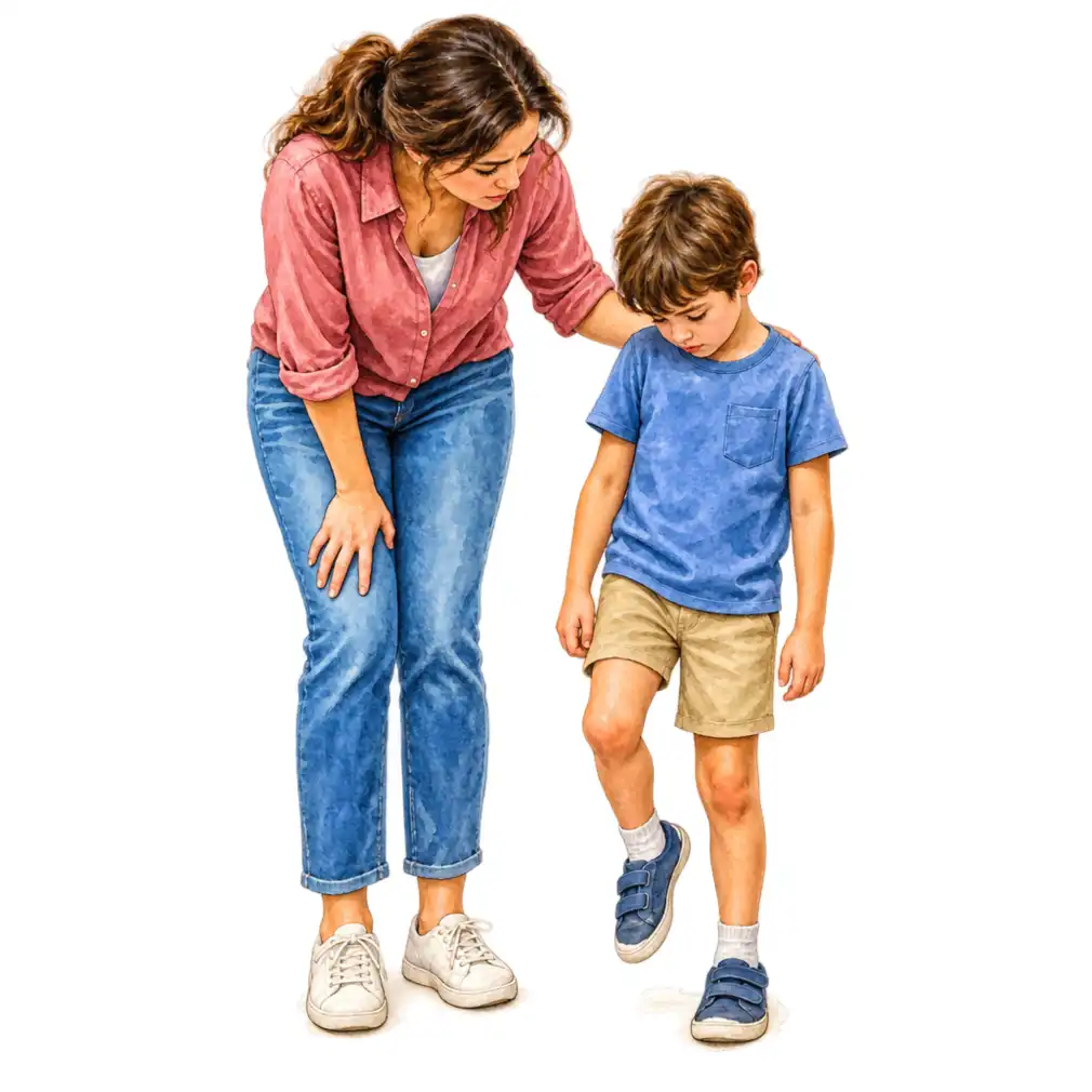
पुरानी चोट के बाद ये संकेत दिखें तो जाँच ज़रूरी हैAfter an old injury, any of these deserves an X-ray
- पैर धीरे-धीरे टेढ़ा होता दिख रहा है — फ्रैक्चर ठीक होने के महीनों बाद, बिना किसी नई चोट के।The limb is slowly bending — months after the fracture healed, with no new injury.
- एक पैर छोटा लग रहा है — खड़े होने पर कूल्हा एक तरफ़ नीचे, या पैंट की एक मोहरी ऊँची।One leg looks shorter — a hip that sits lower on standing, or one trouser leg riding high.
- घुटने या टखने की रेखा तिरछी दिख रही है, या चलते हुए घुटना अंदर/बाहर जा रहा है।A tilted knee or ankle line, or a knee that drifts in or out during walking.
- चोट के बहुत बाद शुरू हुई लंगड़ाहट — जो पहले नहीं थी।A limp that started long after the injury, when there was none before.
- जूते एक तरफ़ से ज़्यादा घिस रहे हैं या बच्चा जल्दी थक जाता है।Shoes wearing down unevenly, or a child tiring quickly on walks.
- कलाई के पास की चोट के बाद हाथ मोड़ने-घुमाने में दिक़्क़त या कलाई का टेढ़ा दिखना।After a wrist injury, a forearm that will not rotate fully or a wrist that looks crooked.
- बचपन में हड्डी/जोड़ का संक्रमण हुआ हो और अब उस अंग की बढ़त पीछे लग रही हो।A history of bone or joint infection in infancy, with that limb now lagging behind.
जाँच कैसे होती है
How It Is Diagnosed
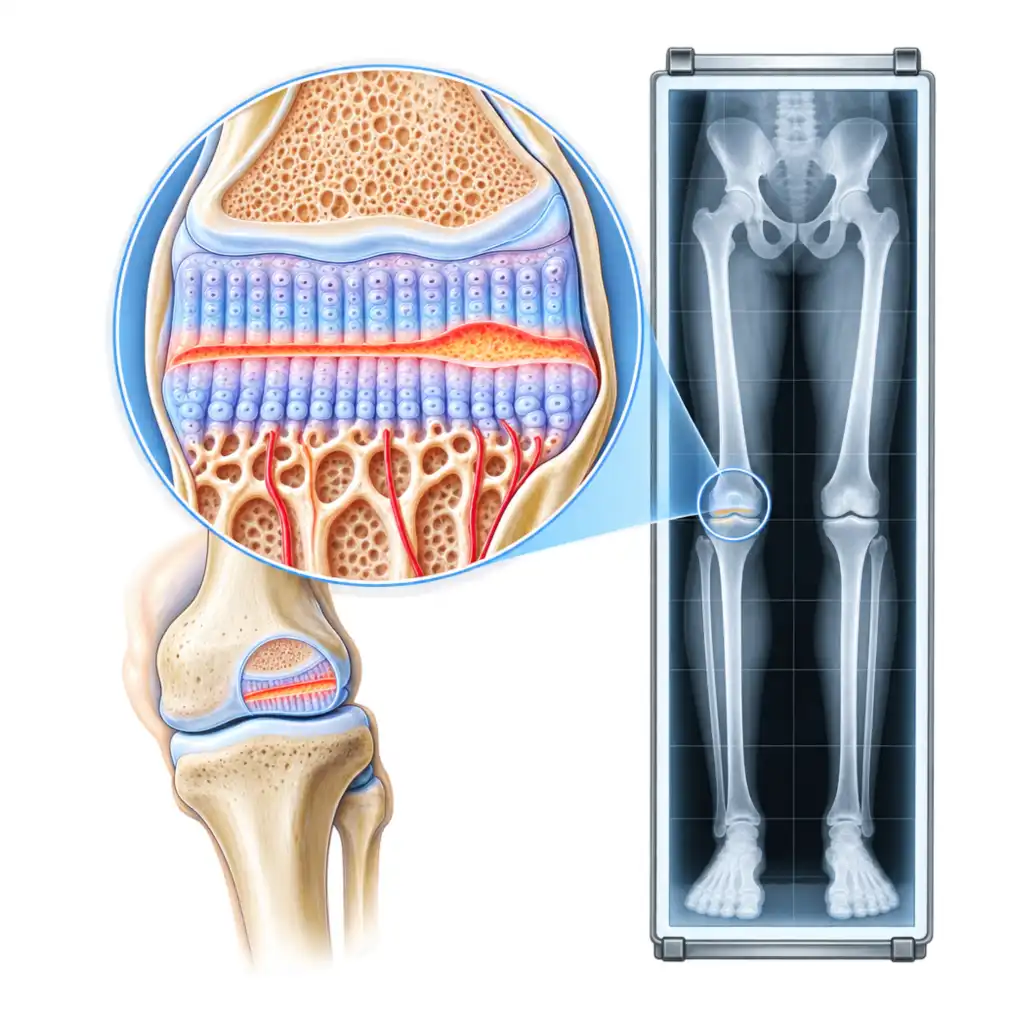
Physeal bar की जाँच में सबसे क़ीमती चीज़ पुरानी X-ray होती है। एक अकेली फ़िल्म बताती है कि आज क्या है; कई फ़िल्में साथ रखकर देखने पर पता चलता है कि यह कितनी तेज़ी से बिगड़ रहा है — और असली फ़ैसला उसी पर टिका होता है।
The most valuable thing you can bring to this consultation is the old X-rays. A single film shows what the bone looks like today; a run of films shows how fast it is going wrong — and that is what the decision actually turns on.
दोनों तरफ़ की X-rayX-rays of both sides
चोट वाली और स्वस्थ — दोनों हड्डियों की तुलना ही सबसे साफ़ जवाब देती है। खड़े होकर ली गई पूरी लंबाई की फ़िल्म (scanogram) से लंबाई और कोण दोनों नापे जाते हैं।Injured side and normal side together, because the comparison is what makes the answer obvious. A standing full-length film measures both the length difference and the angle.
Harris lines — हड्डी की अपनी गवाहीHarris lines - the bone's own evidence
बीमारी या चोट के बाद हड्डी में बनी बारीक सफ़ेद रेखाएँ। ये सामान्यतः growth plate के समानांतर चलती हैं; अगर ये किसी एक बिंदु की ओर झुकती हुई मिलें, तो वहीं bar है।The fine white lines left in bone after an illness or injury normally run parallel to the plate. Where they converge towards one spot, that is where the tether is.
MRI — bar का नक़्शाMRI - mapping the bar
सबसे अहम जाँच। इससे पता चलता है कि bar कहाँ है और पूरी plate का कितने प्रतिशत घेरता है। यही प्रतिशत तय करता है कि इसे निकाला जा सकता है या नहीं।The decisive investigation. It shows where the bar is and what percentage of the plate it occupies — and that percentage decides whether it can be removed at all.
CT — जब हड्डी का आकार देखना होCT - when the shape matters
3D reconstruction से bar का आकार और उस तक पहुँचने का रास्ता साफ़ दिखता है, ख़ासकर जब पहले से कोई implant लगा हो।A 3D reconstruction shows the shape of the bar and the safest route to it, particularly where implants are already in place.
Bone age — बची हुई growthBone age - how much growth is left
बाएँ हाथ की X-ray से हड्डी की उम्र निकाली जाती है। इससे अंदाज़ा लगता है कि आगे कितनी growth बाक़ी है — और यही तय करता है कि bar निकालना फ़ायदेमंद होगा या नहीं।An X-ray of the left hand gives the skeletal age, and from it the growth still to come. That figure is what makes a resection worth doing — or not.
आगे की लंबाई का अनुमानPredicting the final difference
Multiplier और Green-Anderson जैसे तरीक़ों से यह अनुमान लगाया जाता है कि growth पूरी होने पर अंतर कितना होगा — क्योंकि इलाज आज के अंतर पर नहीं, अंतिम अंतर पर तय होता है।Multiplier and Green-Anderson methods estimate what the difference will be at skeletal maturity, because treatment is planned around the final discrepancy, not today's.
📁 पुरानी फ़िल्में फेंके नहीं📁 Do not throw the old films away
चोट के दिन की, plaster खुलने के समय की, और बीच की हर X-ray — तारीख़ के हिसाब से एक फ़ाइल में रखें। मोबाइल में फ़ोटो भी चलेगी। यही सबसे तेज़ी से बताती हैं कि bar बढ़ रहा है या रुका हुआ है।The film from the day of the injury, the one from when the cast came off, and everything in between — keep them in one folder in date order. Phone photographs of the films are fine. Nothing tells us faster whether the bar is doing damage or sitting still.
इलाज — फ़ैसला दो बातों पर टिका है
Treatment - The Decision Rests on Two Numbers
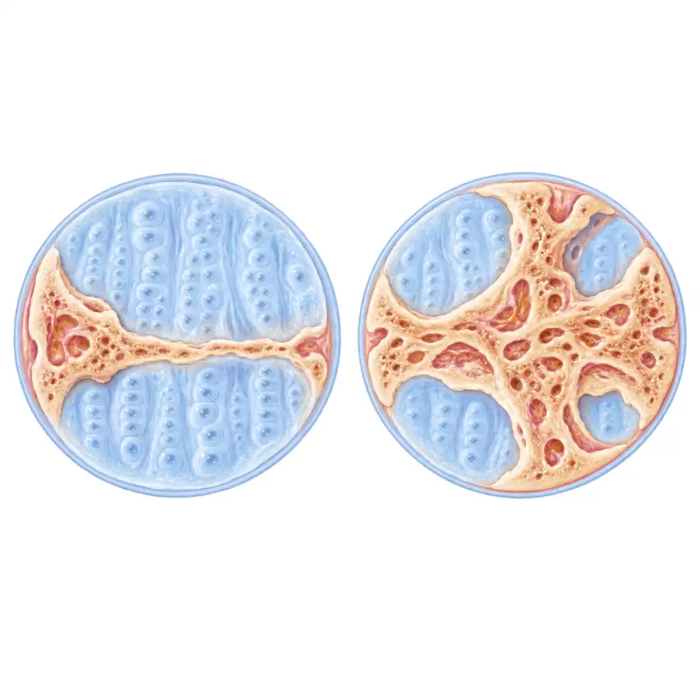
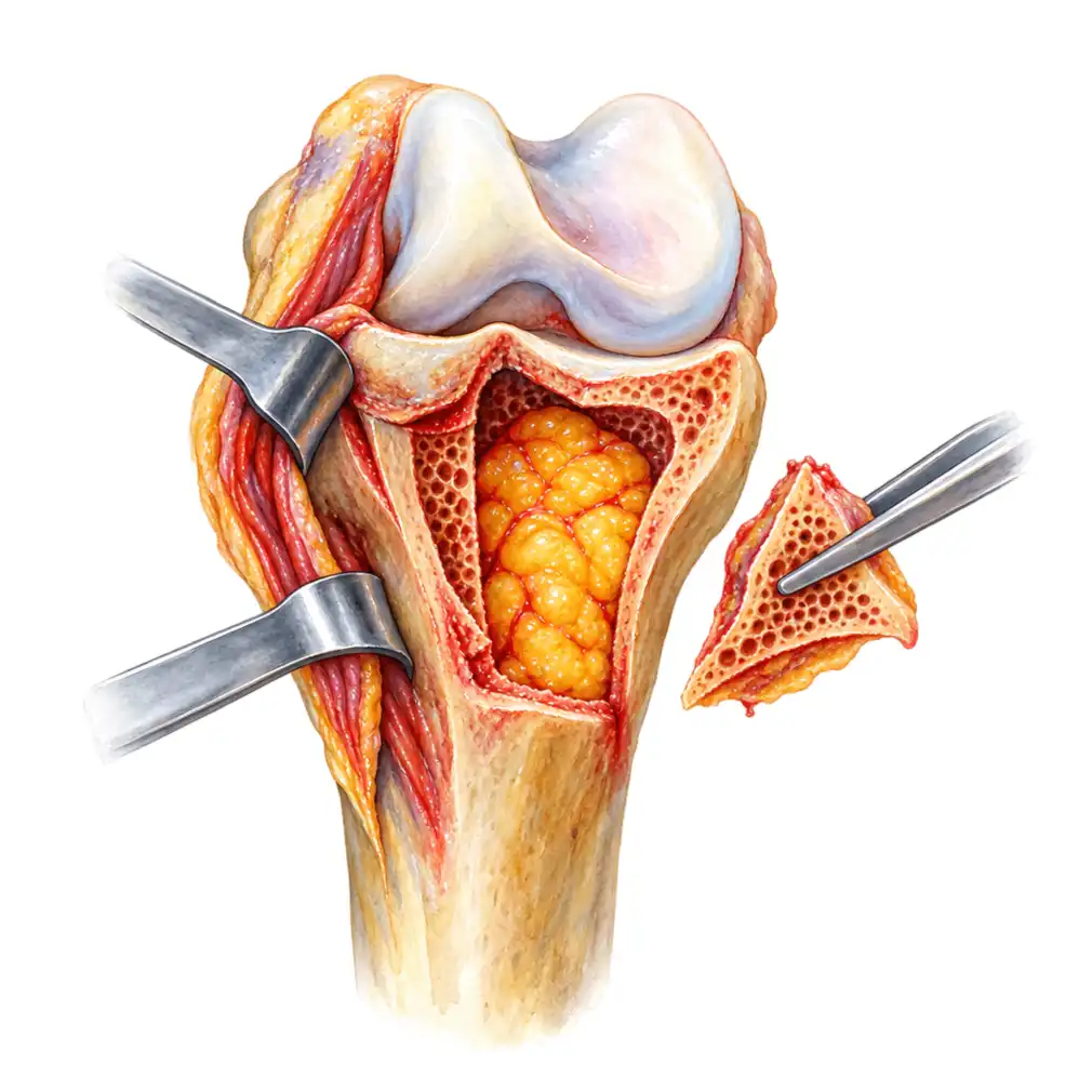
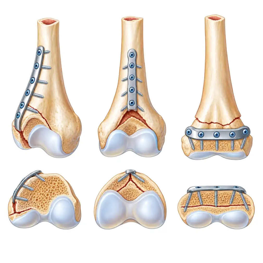
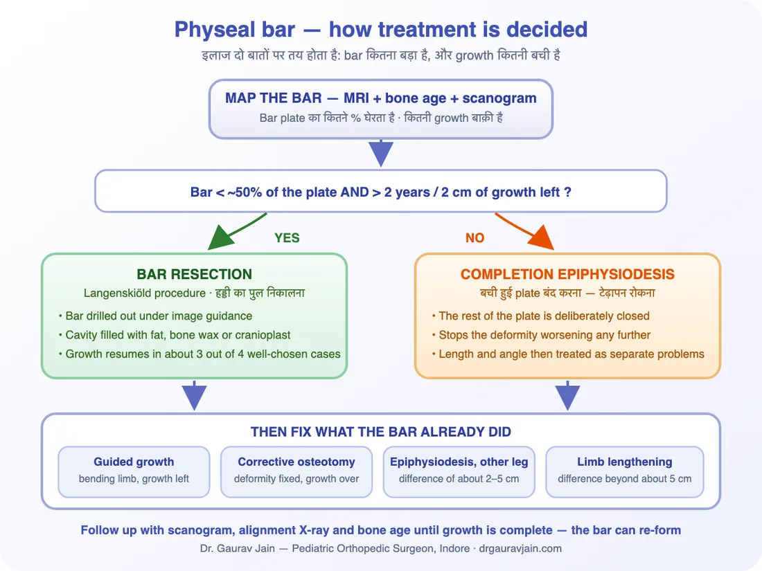
Physeal bar की पूरी सर्जरी दो नंबरों के आसपास घूमती है: bar plate का कितने प्रतिशत घेरता है, और बच्चे की कितनी growth बची है। बाक़ी सब इन्हीं दोनों से निकलता है।
The whole of physeal bar surgery turns on two numbers: what fraction of the plate the bar occupies, and how much growth the child has left. Everything else follows from those two.
| स्थितिSituation | आमतौर पर क्या किया जाता हैWhat is usually done | मक़सदThe aim |
|---|---|---|
| छोटा bar (~50% से कम), 2 साल से ज़्यादा growth बाक़ीSmall bar (under about 50%), more than 2 years of growth left | Bar resection + fat/cranioplast interpositionBar resection with fat or cranioplast interposition | Growth plate को दोबारा चालू करनाRestart the growth plate |
| छोटा bar, पर साथ में साफ़ टेढ़ापनSmall bar, but the limb is already visibly bent | Resection के साथ guided growth या osteotomyResection combined with guided growth or an osteotomy | Growth बचाना और साथ ही सीधा करनाSave the growth and straighten at the same time |
| बड़ा bar (~50% से ज़्यादा) या growth बहुत कम बचीLarge bar (over about 50%) or little growth left | बची हुई plate की completion epiphysiodesisCompletion epiphysiodesis of the remaining plate | टेढ़ापन आगे बढ़ने से रोकनाStop the deformity getting any worse |
| लंबाई का अंतर 2–5 cm रहने का अनुमानPredicted difference of about 2 to 5 cm | सही समय पर दूसरे पैर की epiphysiodesisA well-timed epiphysiodesis on the longer leg | Growth ख़त्म होने तक दोनों पैर बराबरLegs that finish level at maturity |
| अंतर 5 cm से ज़्यादा रहने का अनुमानPredicted difference beyond about 5 cm | Limb lengthening (Ilizarov / rail / lengthening nail)Limb lengthening (Ilizarov, rail fixator or a lengthening nail) | छोटे पैर की लंबाई वापस लानाGive the short limb its length back |
| Growth लगभग पूरी, टेढ़ापन स्थिरGrowth nearly finished, deformity now static | Corrective osteotomyCorrective osteotomy | एक बार में सीधा करनाStraighten it once and for all |
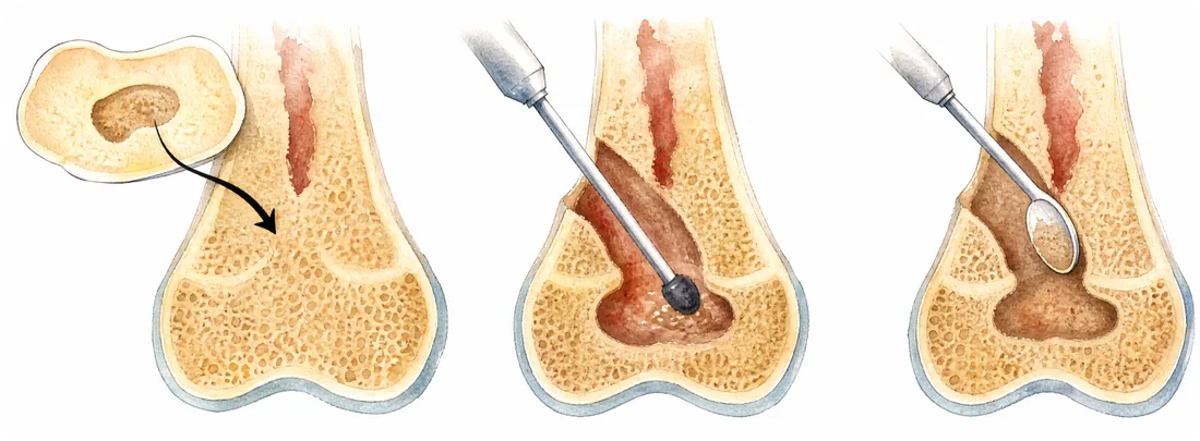
Bar resection (Langenskiöld procedure)Bar resection (Langenskiold procedure)
X-ray guidance में हड्डी का पुल drill करके निकाला जाता है, और बनी हुई जगह में चर्बी (fat), bone wax या cranioplast भर दिया जाता है ताकि हड्डी दोबारा पुल न बना सके। मुक्त हुई plate फिर से बढ़ना शुरू कर देती है।The bridge of bone is drilled away under image guidance and the cavity filled with the child's own fat, bone wax or cranioplast so bone cannot bridge across again. The released plate then resumes growing.
Guided growth (hemiepiphysiodesis)Guided growth (hemiepiphysiodesis)
बची हुई growth का इस्तेमाल करके पैर को सीधा किया जाता है — plate के एक तरफ़ छोटी plate या screw लगाकर उस तरफ़ की बढ़त धीमी कर दी जाती है। हड्डी काटनी नहीं पड़ती।Uses the growth that remains to do the correcting: a small plate or screw slows one side of the physis and the limb straightens itself over months. No bone is cut.
Completion epiphysiodesisCompletion epiphysiodesis
जब bar इतना बड़ा हो कि निकालना बेकार है, तो बची हुई plate को जानबूझकर बंद कर दिया जाता है — ताकि टेढ़ापन और न बढ़े। लंबाई का हिसाब बाद में अलग से किया जाता है।When the bar is too big to be worth removing, the rest of the plate is deliberately closed so the limb cannot bend any further. Length is then dealt with as a separate problem.
दूसरे पैर की epiphysiodesisEpiphysiodesis of the other leg
लंबे पैर की growth plate को सही उम्र पर बंद करके दोनों पैरों को बराबर पर लाया जाता है। छोटा ऑपरेशन, पर timing ही सब कुछ है — इसीलिए bone age बार-बार देखी जाती है।Closing the plate in the longer leg at the calculated moment lets the short leg catch up. A small operation in which timing is everything — hence the repeated bone-age films.
Limb lengtheningLimb lengthening
बड़े अंतर के लिए हड्डी को धीरे-धीरे बढ़ाया जाता है — Ilizarov ring, rail fixator या अंदर लगने वाली lengthening nail से। लंबा रास्ता है, पर नतीजा टिकाऊ।For larger differences the bone is lengthened gradually with an Ilizarov ring, a rail fixator or an internal lengthening nail. A long road, but a durable result.
Corrective osteotomyCorrective osteotomy
जब growth लगभग ख़त्म हो चुकी हो या टेढ़ापन बहुत ज़्यादा हो, तो हड्डी काटकर सही कोण पर जोड़ी जाती है और plate-screw से स्थिर कर दी जाती है।Where growth is nearly over or the deformity is large, the bone is cut, realigned and held with a plate and screws.
🎯 Resection से क्या उम्मीद रखें🎯 What to expect from a resection
सही चुने गए बच्चों में — छोटा bar, अच्छी-ख़ासी growth बाक़ी, बाक़ी plate स्वस्थ — लगभग चार में से तीन मामलों में growth दोबारा शुरू हो जाती है। Bar दोबारा भी बन सकता है, इसलिए growth पूरी होने तक हर साल X-ray चलती रहती है। यह असफलता नहीं, बढ़ती हड्डी का स्वभाव है।In carefully selected children — a small bar, plenty of growth left, an otherwise healthy plate — growth resumes in roughly three out of four. The bar can re-form, so yearly X-rays continue until growth is complete. That is not a failure of the operation; it is what growing bone does.
Operation के बाद — क्या और कब
After Surgery - What Happens When
अस्पताल
Bar निकालकर जगह में fat या cranioplast भरा जाता है। ज़्यादातर बच्चे एक-दो दिन में घर चले जाते हैं। दर्द आमतौर पर कम रहता है।
बचाव के साथ चलना
Crutches या walker के सहारे सीमित वज़न। घाव भरते ही स्कूल शुरू किया जा सकता है।
सामान्य गतिविधि
पूरा वज़न और रोज़ की सारी गतिविधियाँ। जोड़ की movement और muscle strength के लिए physiotherapy।
पहला असली जवाब
X-ray पर देखा जाता है कि growth plate दोबारा चली या नहीं — Harris line अब plate के समानांतर बननी चाहिए। यही सबसे अहम मुलाक़ात है।
Growth ख़त्म होने तक
Scanogram, alignment X-ray और bone age। Bar दोबारा बन सकता है, और लंबाई का अंतर भी बदलता रहता है — इसलिए follow-up बीच में नहीं छोड़ना है।
In hospital
The bar is removed and the cavity filled with fat or cranioplast. Most children go home within a day or two, and pain is usually modest.
Protected walking
Limited weight through the leg with crutches or a walker. School restarts once the wound is settled.
Back to normal activity
Full weight and ordinary daily life, with physiotherapy for joint movement and muscle strength.
The first real answer
An X-ray shows whether the plate has restarted — a new Harris line should now be running parallel to it. This is the visit that matters most.
Until growth ends
Scanograms, alignment films and bone age. The bar can re-form and the length difference keeps changing, so this is not the follow-up to abandon halfway.
रोकथाम और चोट के बाद की निगरानी
Prevention and Follow-Up After a Growth Plate Injury
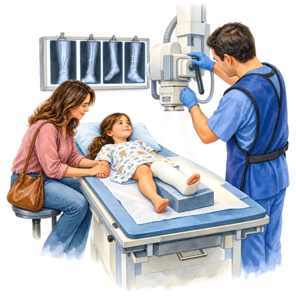
हर bar रोका नहीं जा सकता — कुछ नुक़सान चोट के उसी क्षण हो जाता है। लेकिन ख़तरा वाक़ई कम किया जा सकता है, और जल्दी पकड़ में आ जाना तो पूरी तरह हमारे हाथ में है।
Not every bar can be prevented — some of the damage is done in the instant of the injury. But the risk can genuinely be lowered, and catching it early is entirely within our control.
- पहली बार में सही reduction — growth plate की चोट को जल्दी और सही तरीक़े से बैठाना सबसे बड़ी सुरक्षा है।An accurate reduction the first time. Getting a physeal fracture into position early and properly is the single biggest protection.
- 7–10 दिन बाद बार-बार ज़ोर लगाकर सीधा करने से बचना — इससे plate को और चोट पहुँचती है।No repeated forceful manipulation after the first week to ten days — each attempt adds injury to the plate.
- Implant का रास्ता सोच-समझकर — हो सके तो screw और wire growth plate को पार न करें, और समय पर निकाल दिए जाएँ।Thoughtful implant placement — wires and screws kept out of the plate wherever possible, and removed on time.
- बच्चों में हड्डी/जोड़ के संक्रमण को आपात स्थिति मानना — देर से इलाज़ हुआ septic arthritis growth plate को स्थायी नुक़सान पहुँचा सकता है।Treating bone and joint infection in children as an emergency — a late-treated septic joint can take the growth plate with it.
- Discharge के समय परिवार को साफ़ बताना कि यह growth plate की चोट थी और आगे किन बातों पर नज़र रखनी है।Telling the family plainly at discharge that this was a growth plate injury, and exactly what to watch for afterwards.
- Follow-up बीच में न छोड़ना — यह अकेली आदत सबसे ज़्यादा बच्चों को बड़ी सर्जरी से बचाती है।Not abandoning follow-up. This one habit spares more children a major operation than anything else on the list.
Growth plate की चोट के बाद जाँच का कार्यक्रमThe follow-up schedule after a growth plate injury
✅ जल्दी पकड़ में आने का फ़ायदा साफ़ है✅ Why catching it early changes everything
छोटा bar निकाला जा सकता है और growth वापस आ सकती है। वही bar दो साल बाद बड़ा हो चुका होता है, पैर टेढ़ा हो चुका होता है, और तब osteotomy या lengthening जैसी बड़ी सर्जरी की ज़रूरत पड़ती है। फ़र्क़ बीमारी में नहीं — समय में है।A small bar can be removed and the growth recovered. The same bar two years later is bigger, the limb is bent, and the answer becomes an osteotomy or a lengthening. The difference is not in the disease — it is in when it was found.
भ्रम बनाम सच्चाई
Myths vs Facts
"फ्रैक्चर जुड़ गया, अब कोई ख़तरा नहीं।"
हड्डी जुड़ना और growth plate का बचना दो अलग बातें हैं। फ्रैक्चर पूरी तरह ठीक होने के बाद भी plate पर पुल बन सकता है — और असर महीनों बाद दिखता है।
"दर्द नहीं है, तो कुछ गड़बड़ नहीं होगी।"
Physeal bar लगभग हमेशा बिना दर्द के बढ़ता है। इसे दर्द नहीं, बदलती हुई शक्ल पकड़वाती है।
"Calcium, विटामिन और अच्छे खाने से growth plate दोबारा चल पड़ेगी।"
Vitamin D की कमी का इलाज अपनी जगह ज़रूरी है, पर जो हड्डी का पुल बन चुका है उसे किसी दवा या खुराक से नहीं हटाया जा सकता।
"बच्चा बड़ा होगा तो टेढ़ापन अपने आप सीधा हो जाएगा।"
यहाँ उल्टा होता है — बच्चा जितना बढ़ेगा, tether उतना ही ज़्यादा टेढ़ा करेगा। इंतज़ार से हालत सुधरती नहीं, बिगड़ती है।
"ऑपरेशन का नतीजा तुरंत पता चल जाएगा।"
नतीजा growth से नापा जाता है। पहला भरोसेमंद जवाब 6 महीने की X-ray पर मिलता है, पूरा जवाब growth ख़त्म होने पर।
"मालिश और कसरत से पैर सीधा हो जाएगा।"
Physiotherapy जोड़ की movement और ताक़त के लिए ज़रूरी है, पर यह हड्डी के कोण को नहीं बदल सकती — वह हड्डी के अंदर की समस्या है।
"The fracture united, so we are past all this."
Union of the bone and survival of the growth plate are two separate questions. A bar can form across a perfectly healed fracture, and only declares itself months later.
"There is no pain, so nothing can be wrong."
A physeal bar is almost always painless. What gives it away is not pain but a shape that is quietly changing.
"Calcium, vitamins and good food will restart the growth plate."
A vitamin D deficiency must be corrected on its own merits, but no supplement dissolves a bridge of bone that has already formed.
"He will straighten out as he grows."
Here the opposite is true: the more the child grows, the more the tether bends the bone. Waiting does not improve this — it worsens it.
"We will know straight away whether the operation worked."
The result is measured in growth. The first reliable answer is on the six-month X-ray, the final one when growth ends.
"Massage and exercises will straighten the leg."
Physiotherapy is important for joint movement and strength, but it cannot change the angle of a bone. That problem sits inside the bone.
इससे जुड़ी बातें
Related Reading
Physeal bar अकेले नहीं आता — यह अक्सर किसी पुरानी चोट का नतीजा होता है और आगे चलकर लंबाई या टेढ़ेपन की समस्या बन जाता है। इन पन्नों पर वही आगे-पीछे की कहानी है।
A physeal bar rarely arrives alone — it follows an old injury and it leads to a length or alignment problem. These pages cover what came before and what comes after.
Indore में जाँच — क्या साथ लाएँ
Getting Checked in Indore - What to Bring
Physeal bar में सबसे महँगी चीज़ खोया हुआ समय है। Dr. Gaurav Jain (MBBS, D.Ortho, DNB Orthopaedics, MNAMS; बच्चों की हड्डी रोग और स्पाइन सर्जरी में फेलोशिप) Indore में बच्चों की growth plate की चोट, growth arrest, पैरों की लंबाई के अंतर और हड्डी की विकृति का इलाज करते हैं — bar resection, guided growth, epiphysiodesis, corrective osteotomy और limb lengthening सहित। Bhopal में Rainbow Children Hospital में वैकल्पिक रविवार को भी परामर्श उपलब्ध है।
In a physeal bar the most expensive thing is lost time. Dr. Gaurav Jain (MBBS, D.Ortho, DNB Orthopaedics, MNAMS; fellowship-trained in pediatric orthopedics and spine surgery) treats growth plate injury, growth arrest, limb length discrepancy and deformity in children in Indore — bar resection, guided growth, epiphysiodesis, corrective osteotomy and limb lengthening. Consultations are also available at Rainbow Children Hospital, Bhopal, on alternate Sundays.
- चोट के दिन से अब तक की सारी X-ray, तारीख़ के हिसाब से — तुलना ही सबसे ज़्यादा बताती है
- अस्पताल के काग़ज़ — discharge summary, operation notes, और वह पर्ची जिस पर Salter-Harris type लिखा हो
- पहले हो चुकी MRI या CT, फ़िल्म और रिपोर्ट दोनों
- बच्चे की लंबाई का रिकॉर्ड और, अगर हो तो, पैरों की लंबाई की पुरानी माप
- खड़े होकर और चलते हुए का video — सामने और पीछे दोनों से
- संक्रमण या दूसरी सर्जरी हुई हो तो उसके काग़ज़
- Every X-ray from the day of the injury onwards, in date order — the comparison tells us the most
- The hospital paperwork — discharge summary, operation notes, and the slip that names the Salter-Harris type
- Any MRI or CT already done, both the films and the report
- The child's height records and any earlier leg-length measurements
- A video standing and walking, from in front and from behind
- Notes from any infection or previous surgery
अक्सर पूछे जाने वाले सवाल
Frequently Asked Questions
Physeal bar क्या है? / What is a physeal bar?
Growth plate — यानी वह cartilage की पट्टी जिससे बच्चे की हड्डी लंबी होती है — पर बन जाने वाला हड्डी का पुल। जहाँ पुल है वहाँ बढ़त रुक जाती है, बाक़ी plate बढ़ती रहती है, इसलिए हड्डी या तो टेढ़ी होती जाती है या बढ़ना बंद कर देती है।
A bridge of solid bone across the growth plate — the band of cartilage from which a child's bone lengthens. Wherever the bridge sits growth stops while the rest of the plate carries on, so the bone either bends progressively or stops lengthening altogether.
फ्रैक्चर तो ठीक हो गया था, अब पैर क्यों टेढ़ा हो रहा है? / The fracture healed — why is the leg bending now?
क्योंकि फ्रैक्चर जुड़ना और growth plate बचना दो अलग बातें हैं। हड्डी पूरी तरह जुड़ सकती है और फिर भी plate पर पुल बन सकता है। यह पुल तभी नुक़सान करता है जब बच्चा बढ़ता है — इसीलिए टेढ़ापन चोट के 6 महीने से 2 साल बाद दिखना शुरू होता है।
Because union of the fracture and survival of the growth plate are different things. The bone can heal perfectly and still leave a bar behind, and the bar only does harm as the child grows — which is why the bending appears six months to two years after the injury.
कौन-सी चोट के बाद ख़तरा ज़्यादा है? / Which injuries carry the most risk?
Salter-Harris type III और IV (जो plate को पार करके जोड़ तक जाती हैं) और type V (plate का कुचल जाना) सबसे ख़तरनाक हैं। जगह के हिसाब से घुटने के ठीक ऊपर की चोट सबसे बदनाम है — यहाँ सही reduction के बाद भी growth बिगड़ सकती है।
Salter-Harris types III and IV, which cross the plate into the joint, and type V crush injuries. By location, the fracture just above the knee is the most notorious — growth can be disturbed there even after an excellent reduction.
क्या इसमें दर्द होता है? / Does a physeal bar hurt?
आमतौर पर नहीं — और यही सबसे बड़ी दिक़्क़त है। ज़्यादातर बच्चे इसलिए लाए जाते हैं क्योंकि माता-पिता ने पैर टेढ़ा होते या जूते एक तरफ़ से घिसते देखा, दर्द की शिकायत के कारण नहीं।
Usually not, and that is precisely the problem. Most children are brought in because a parent noticed the limb bending or the shoes wearing unevenly, not because anything hurts.
क्या संक्रमण से भी bar बन सकता है? / Can infection cause a bar too?
हाँ। शिशु अवस्था में osteomyelitis या septic arthritis growth plate का हिस्सा नष्ट कर सकता है और पीछे bar छोड़ सकता है। ऐसे बच्चों में विकृति ज़्यादा गंभीर होती है क्योंकि बढ़ने के लिए पूरी उम्र बाक़ी होती है — इसलिए इनकी growth skeletal maturity तक देखी जानी चाहिए।
Yes. Osteomyelitis or septic arthritis in infancy can destroy part of the plate and leave a bar. The deformities are often severe because so much growth remains, so any child treated for a bone or joint infection should have growth followed to skeletal maturity.
जाँच कैसे होती है? / How is it diagnosed?
दोनों तरफ़ की X-ray से शुरुआत, जहाँ Harris lines देखी जाती हैं — ये सामान्यतः plate के समानांतर चलती हैं, और किसी बिंदु की ओर झुकती दिखें तो वहीं bar है। इसके बाद MRI से पता चलता है कि bar कहाँ है और plate का कितने प्रतिशत घेरता है। साथ में bone age और scanogram।
It begins with X-rays of both sides, looking at the Harris growth arrest lines — normally parallel to the plate, and converging on the tether where a bar has formed. An MRI then maps where the bar is and what fraction of the plate it takes up, alongside a bone age and a scanogram.
MRI क्यों ज़रूरी है, X-ray काफ़ी नहीं? / Why an MRI - is an X-ray not enough?
X-ray बता देती है कि गड़बड़ है, पर कितनी है यह नहीं। इलाज का पूरा फ़ैसला इस बात पर है कि bar plate का कितने प्रतिशत घेरता है — और वह नाप सिर्फ़ MRI (या कभी CT) पर मिलती है।
An X-ray tells us that something is wrong; it does not tell us how much. The entire treatment decision rests on what percentage of the plate the bar occupies, and only an MRI — occasionally a CT — gives that measurement.
क्या growth plate ठीक की जा सकती है? / Can the growth plate be repaired?
Bar निकाला जा सकता है। Bar resection में हड्डी का पुल drill करके हटाया जाता है और खाली जगह में चर्बी, bone wax या cranioplast भर दिया जाता है ताकि पुल दोबारा न बने। यह तब सबसे कारगर है जब bar plate के लगभग आधे से कम हो और बच्चे की कम से कम 2 साल या 2 cm growth बाक़ी हो।
The bar can be removed. In a bar resection the bridge is drilled out and the cavity filled with fat, bone wax or cranioplast so bone cannot bridge again. It works best when the bar covers less than about half the plate and the child has at least two years or two centimetres of growth still to come.
अगर bar बहुत बड़ा हो तो? / What if the bar is too large?
तब मक़सद बदल जाता है — growth बचाने के बजाय नतीजे को क़ाबू में रखना। बची हुई plate जानबूझकर बंद कर दी जाती है ताकि टेढ़ापन और न बढ़े, विकृति guided growth या osteotomy से सुधारी जाती है, और लंबाई का हिसाब दूसरे पैर की epiphysiodesis या lengthening से किया जाता है।
The aim changes from saving growth to controlling the outcome. The remaining plate is deliberately closed so the limb cannot bend further, the deformity is corrected by guided growth or an osteotomy, and length is balanced by epiphysiodesis of the other leg or by lengthening.
Guided growth कब काम आता है? / When does guided growth help?
जब growth बाक़ी हो और समस्या टेढ़ेपन की हो, लंबाई की नहीं। Plate के स्वस्थ हिस्से पर छोटी plate या screw लगाकर उस तरफ़ की बढ़त धीमी कर दी जाती है और पैर महीनों में ख़ुद सीधा हो जाता है — हड्डी काटे बिना।
When growth remains and the problem is a bend rather than a shortening. A small plate or screw on the healthy side of the physis slows it, and the limb straightens itself over months without any bone being cut.
क्या limb lengthening करानी पड़ेगी? / Will my child need limb lengthening?
सिर्फ़ तब, जब अनुमानित अंतर इतना बड़ा हो कि दूसरे पैर को धीमा करके संभाला न जा सके — आमतौर पर लगभग 5 cm से ज़्यादा। इससे कम अंतर सही समय पर की गई epiphysiodesis से संभल जाता है, और बहुत छोटा अंतर जूते में raise से।
Only when the predicted difference is beyond what slowing the other leg can absorb — usually more than about five centimetres. Smaller differences are handled with a well-timed epiphysiodesis, and very small ones with a shoe raise.
ठीक होने में कितना समय लगता है? / How long is the recovery?
Bar resection के बाद 2–4 हफ़्ते बचाव के साथ चलना और आमतौर पर 2–4 हफ़्ते में स्कूल वापसी; 6–8 हफ़्ते में सामान्य गतिविधि। लेकिन ऑपरेशन का असली नतीजा घाव भरने से नहीं, growth से नापा जाता है — पहला भरोसेमंद जवाब 6 महीने की X-ray पर मिलता है।
After a bar resection, two to four weeks of protected walking, school back within two to four weeks and normal activity by six to eight. But the operation's real result is measured in growth, not healing — the first reliable answer comes on the six-month X-ray.
Growth plate की चोट के बाद कितने समय तक दिखाना चाहिए? / How long is follow-up needed after a growth plate fracture?
कम से कम 1 साल, और घुटने के आसपास की ख़तरनाक चोट के बाद 2 साल — यानी उस समय से काफ़ी आगे तक जब बच्चा बिल्कुल ठीक महसूस कर रहा हो। 3, 6, 12 और 24 महीने की जाँच लगभग मुफ़्त है और bar को तब पकड़ लेती है जब वह अभी छोटा और निकालने लायक़ है।
At least one year, and two years after a high-risk injury around the knee — well past the point where the child feels completely fine. Checks at three, six, twelve and twenty-four months cost almost nothing and catch a bar while it is still small enough to remove.
Bar resection कितनी कामयाब होती है? / How successful is bar resection?
सही चुने गए बच्चों में — छोटा bar, अच्छी-ख़ासी growth बाक़ी, बाक़ी plate स्वस्थ — लगभग चार में से तीन में growth दोबारा शुरू हो जाती है। Bar दोबारा बन सकता है, इसलिए growth ख़त्म होने तक हर साल X-ray ज़रूरी है, और कभी-कभी दूसरी प्रक्रिया की ज़रूरत पड़ती है।
In well-selected children — a small bar, plenty of growth left, an otherwise healthy plate — growth resumes in roughly three out of four. The bar can re-form, so yearly X-rays continue until growth ends, and a second procedure is occasionally needed.
क्या इसे रोका जा सकता है? / Can a physeal bar be prevented?
पूरी तरह नहीं, क्योंकि कुछ नुक़सान चोट के उसी क्षण हो जाता है। पर ख़तरा वाक़ई कम होता है — पहली बार में सही reduction से, 7–10 दिन बाद बार-बार ज़ोर लगाकर सीधा करने से बचकर, implant को plate से दूर रखकर, और बच्चों में हड्डी-जोड़ के संक्रमण को आपात स्थिति मानकर।
Not entirely, because some of the damage happens at the moment of injury. But the risk genuinely falls with an accurate early reduction, by avoiding repeated forceful manipulation after the first week, by keeping implants out of the plate, and by treating children's bone and joint infections as emergencies.
क्या बच्चा आगे खेल-कूद पाएगा? / Will my child play sport again?
हाँ। Bar resection के बाद घाव भरते ही सामान्य गतिविधि लौट आती है, और कोण व लंबाई ठीक हो जाने पर ज़्यादातर बच्चे बिना किसी रोक-टोक के दौड़ते-खेलते हैं। Contact sport कब शुरू करना है, यह सर्जन तय करते हैं।
Yes. Normal activity returns once the wound has settled after a resection, and with the angle and length corrected most children run and play without restriction. The timing of contact sport is your surgeon's call.
Indore में किसे दिखाएँ? / Which doctor should we see in Indore?
एक pediatric orthopedic surgeon, क्योंकि यहाँ फ़ैसला अकेली X-ray से नहीं, बल्कि बची हुई growth से होता है। Indore में Dr. Gaurav Jain (MBBS, D.Ortho, DNB Orthopaedics, MNAMS; बच्चों की हड्डी रोग और स्पाइन सर्जरी में फेलोशिप) physeal bar, growth arrest, पैरों की लंबाई का अंतर और विकृति सुधार का इलाज करते हैं।
A pediatric orthopedic surgeon, because the decision depends on growth remaining rather than on the X-ray alone. In Indore, Dr. Gaurav Jain (MBBS, D.Ortho, DNB Orthopaedics, MNAMS; fellowship-trained in pediatric orthopedics and spine surgery) treats physeal bars, growth arrest, limb length discrepancy and deformity correction in children.
📞 पुरानी चोट के बाद पैर टेढ़ा या छोटा लग रहा है? आज ही दिखाएँ
📞 A limb bending or shortening after an old injury? Get it checked now
Indore के Pediatric Orthopedic Surgeon | Growth plate की चोट, bar resection, deformity correction और limb lengtheningPediatric Orthopedic Surgeon in Indore | Growth plate injury, bar resection, deformity correction and limb lengthening
📞 अभी Call करें 📞 Call Nowचोट के दिन से अब तक की सारी X-ray साथ लाएँ — छोटा bar निकाला जा सकता है, बड़ा नहीं। यहाँ देर ही सबसे महँगी पड़ती है।Bring every X-ray since the injury - a small bar can be removed, a large one cannot. Here, delay is the expensive part.