Best Exostosis (Osteochondroma) Treatment in Indore (हड्डी की गाँठ)
Exostosis & Hereditary Multiple Exostoses
Osteochondroma - बच्चों का सबसे common benign bone tumour | ज़्यादातर में सिर्फ़ निगरानी, surgery नहीं | Indore
Osteochondroma - the commonest benign bone tumour of childhood | Most need watching, not surgery | Indore
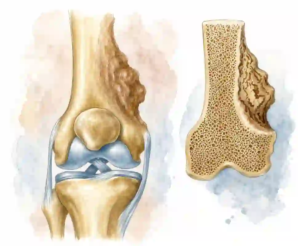
Exostosis क्या है?
What is an Exostosis?
Exostosis (medical नाम: Osteochondroma) हड्डी की एक गाँठ या उभार है जो हड्डी से ही निकलती है और जिसके ऊपर cartilage की टोपी (cap) होती है। यह cancer नहीं है — यह बच्चों का सबसे common benign (हानिरहित) bone tumour है।
🎯 सबसे ज़रूरी बात
यह गाँठ growth plate (हड्डी की बढ़ने वाली प्लेट) के पास से निकलती है और हमेशा joint से दूर की ओर बढ़ती है। यह तभी तक बढ़ती है जब तक बच्चा बढ़ रहा है — puberty के बाद growth रुक जाती है। ज़्यादातर बच्चों में सिर्फ़ निगरानी (observation) चाहिए, operation नहीं।
🦴 यह बनती कैसे है?
Growth plate की थोड़ी सी cartilage हड्डी के किनारे से बाहर खिसक जाती है और वहीं अपनी जगह बढ़ने लगती है। नीचे से यह असली हड्डी में बदलती जाती है और ऊपर cartilage cap बनी रहती है — इसलिए X-ray पर गाँठ का cortex और marrow दोनों मुख्य हड्डी से जुड़े हुए दिखते हैं। यही इसकी पहचान है, और यही इसे किसी खतरनाक tumour से अलग करती है।
An exostosis — its medical name is osteochondroma — is a bony bump that grows out of a bone and carries a cap of cartilage on top. It is not cancer. It is the commonest benign bone tumour in children.
🎯 The one thing to remember
An exostosis grows out of the metaphysis, next to the growth plate, and always points away from the nearest joint. It grows only while your child is growing and stops when the growth plates close after puberty. Most children need watching, not an operation.
🦴 How does it form?
A small piece of growth-plate cartilage slips sideways past the edge of the bone and carries on growing where it lands. Underneath, it steadily turns into real bone while the cartilage cap stays on top. That is why an X-ray shows the lump's cortex and marrow running straight into the parent bone — the single finding that identifies an osteochondroma and separates it from something sinister.
एक गाँठ या कई? — Solitary बनाम Hereditary Multiple Exostoses
One Lump or Many? — Solitary vs Hereditary Multiple Exostoses
यह सबसे पहला सवाल है जिसका जवाब देना ज़रूरी है, क्योंकि दोनों का follow-up और family के लिए मतलब अलग-अलग होता है।
This is the first question worth answering, because the follow-up and what it means for the rest of the family are different in each case.
🔹 Solitary OsteochondromaSolitary Osteochondroma
अकेली गाँठ — सबसे आम स्थिति A single lump — the common situation- सिर्फ़ एक हड्डी पर एक गाँठ
- वंशानुगत (inherited) नहीं — भाई-बहनों को खतरा नहीं
- अक्सर घुटने या कंधे के पास, बिना दर्द के मिलती है
- Cancer में बदलने का खतरा 1% से भी कम
- ज़्यादातर सिर्फ़ observation; तकलीफ़ हो तो excision
- A single lump on one bone
- Not inherited — brothers and sisters are not at risk
- Usually found near the knee or shoulder, painlessly
- Risk of turning cancerous is under 1%
- Usually observed; removed only if it causes trouble
🧬 Hereditary Multiple Exostoses (HME)Hereditary Multiple Exostoses (HME)
कई गाँठें — आनुवंशिक (genetic) स्थिति Many lumps — an inherited condition- कई हड्डियों पर कई गाँठें (MHE / diaphyseal aclasis भी कहते हैं)
- EXT1 या EXT2 gene में बदलाव; autosomal dominant
- माता-पिता में से एक को हो तो हर बच्चे को 50% chance
- ज़्यादातर बच्चों में 12 साल की उम्र से पहले पता चल जाता है
- हड्डी टेढ़ी होना, forearm छोटा रहना, लंबाई में फ़र्क़ — इन पर नज़र ज़रूरी
- वयस्क होने पर cancer में बदलने का खतरा लगभग 1–5% बताया गया है
- Many lumps on several bones (also called MHE or diaphyseal aclasis)
- Caused by a change in the EXT1 or EXT2 gene; autosomal dominant
- If one parent has it, each child has a 50% chance
- Most children are diagnosed before the age of 12
- Bone bowing, a short forearm and limb length difference need watching
- Reported risk of change to cancer in adult life is about 1–5%
ये गाँठें कहाँ बनती हैं?
Where Do Exostoses Grow?
ये वहीं बनती हैं जहाँ हड्डी सबसे तेज़ी से बढ़ती है — यानी लंबी हड्डियों के सिरों (metaphysis) पर। घुटने के आसपास सबसे ज़्यादा।
They appear where bone grows fastest — at the flared ends (metaphyses) of long bones. The area around the knee is by far the commonest.
⚠️ एक बात ध्यान रखेंWorth knowing
उँगलियों की छोटी हड्डियों, pelvis और रीढ़ के पास की गाँठें कम आम हैं, और वयस्क उम्र में इन जगहों पर बदलाव को ज़्यादा गंभीरता से देखा जाता है। इन जगहों की गाँठों के लिए specialist की राय ज़रूरी है। Lumps in the pelvis, around the spine and in the shoulder girdle are less common, and change in these deep sites is taken more seriously in adult life. Lumps in these locations deserve a specialist opinion.
माता-पिता को क्या दिखता है? — लक्षण
What Parents Notice — Symptoms
सबसे आम शुरुआत यही होती है: नहलाते या खेलते समय अचानक एक सख़्त गाँठ हाथ लग जाती है, जो हिलती नहीं — क्योंकि वह हड्डी का ही हिस्सा है।
- सख़्त, हड्डी जैसी गाँठ जो दबाने पर खिसकती नहीं — अक्सर बिना दर्द के
- रगड़ से दर्द — गाँठ के ऊपर से tendon या muscle गुज़रे तो खेलने के बाद दर्द
- गाँठ के ऊपर सूजन (bursa) — बार-बार रगड़ से पानी की थैली बन जाना
- जोड़ का movement कम होना — जैसे घुटना पूरा मोड़ने में रुकावट
- सुन्नपन, झुनझुनी या कमज़ोरी — गाँठ नस (nerve) पर दबाव डाल रही हो
- Forearm छोटा या टेढ़ा दिखना, कलाई का झुकाव — HME में आम
- दोनों पैरों की लंबाई में फ़र्क़ या घुटनों का आपस में टकराना (knock knee)
- अचानक तेज़ दर्द — पतली डंडी वाली गाँठ का टूट जाना (कम आम)
It nearly always starts the same way: while bathing or playing, a parent's hand finds a hard lump that does not move — because it is part of the bone itself.
- A hard, bony lump that does not slide under the skin — usually painless
- Pain from rubbing — when a tendon or muscle rides over the lump, typically after activity
- Swelling over the lump — a bursa formed by repeated friction
- Reduced joint movement — for example a knee that will not fully bend
- Numbness, tingling or weakness — the lump pressing on a nerve
- A short or bowed forearm and a tilted wrist — common in HME
- Unequal leg lengths or knees that knock together
- Sudden sharp pain — a thin stalked lump has fractured (uncommon)
ये संकेत दिखें तो तुरंत दिखाएँ
Warning Signs That Need Prompt Review
Exostosis benign है — पर बहुत कम मामलों में, ख़ासकर बड़ी उम्र में, cartilage cap में बदलाव आ सकता है (chondrosarcoma)। नीचे दिए संकेतों का मतलब यह नहीं कि cancer है; इसका मतलब है कि जाँच करवाना ज़रूरी है।
🚨 इनमें से कुछ भी दिखे तो देर न करें
- बच्चे की growth रुकने के बाद भी गाँठ बढ़ रही हो — यह सबसे अहम संकेत है
- किसी भी उम्र में गाँठ का तेज़ी से बड़ा होना
- आराम करते समय या रात में दर्द, जो activity से जुड़ा न हो
- गाँठ का गर्म, लाल या छूने पर दुखने लगना
- नया सुन्नपन, झुनझुनी, कमज़ोरी या नब्ज़ में फ़र्क़
- MRI पर cartilage cap का मोटा होना — वयस्कों में लगभग 2 cm से ज़्यादा
Perspective ज़रूरी है: अकेली exostosis में यह बदलाव 1% से भी कम होता है, और HME में लगभग 1–5%। यानी 95% से ज़्यादा लोगों में ऐसा कभी नहीं होता। नियमित check-up ही काफ़ी है — डर की ज़रूरत नहीं।
An exostosis is benign — but in a small number of people, usually in adult life, the cartilage cap can change into a low-grade cancer called chondrosarcoma. The signs below do not mean cancer; they mean it needs to be looked at.
🚨 Do not wait if you notice any of these
- A lump that keeps growing after your child has stopped growing — the single most important sign
- Rapid enlargement at any age
- Pain at rest or at night that is not related to activity
- A lump that becomes warm, red or tender
- New numbness, tingling, weakness or a change in the pulse below the lump
- A thickening cartilage cap on MRI — over roughly 2 cm in an adult
Keep the numbers in perspective: this change happens in under 1% of solitary exostoses and a reported 1–5% of people with HME. More than 95 in 100 never face it. Regular review is enough — fear is not required.
HME में हड्डियों का टेढ़ापन — जिस पर नज़र रखनी है
Bone Deformity in HME — What We Watch For
HME में असली चुनौती अक्सर गाँठ खुद नहीं, बल्कि यह होती है कि गाँठें हड्डी की growth को टेढ़ा कर देती हैं। इन्हें समय पर पकड़ लिया जाए तो छोटे-से operation से सुधार हो जाता है।
In HME the real challenge is often not the lump itself but the way lumps pull a growing bone out of shape. Caught in time, these can be corrected with small, well-timed procedures.
छोटा ulna, मुड़ा हुआ radiusShort ulna, bowed radius
Forearm की सबसे आम समस्या। Ulna की growth जल्दी रुक जाती है, radius बढ़ता रहता है — forearm छोटा और टेढ़ा, कलाई झुकी हुई। कभी-कभी radial head अपनी जगह से खिसक जाता है। The commonest forearm problem. The ulna stops growing early while the radius keeps going, so the forearm becomes short and bowed and the wrist tilts. The radial head can slip out of place.
Ankle valgus (टखना बाहर झुकना)Ankle valgus
Fibula की growth पीछे रह जाने से टखना बाहर की ओर झुक जाता है — जूते घिसना, चलने में थकान। Guided growth से आसानी से ठीक होता है। When the fibula lags behind, the ankle tilts outwards — shoes wear unevenly and walking tires the child. Guided growth corrects this well.
Knock knee (घुटनों का टकराना)Knock knee
घुटने के पास की गाँठें growth plate को असमान रूप से खींचती हैं, जिससे टाँगें अंदर की ओर झुक जाती हैं। Lumps beside the knee pull unevenly on the growth plate, and the legs angle inwards.
लंबाई में फ़र्क़ (LLD)Limb length difference
एक टाँग दूसरी से छोटी रह सकती है। 2 cm से ज़्यादा फ़र्क़ हो तो lengthening या epiphysiodesis की planning की जाती है। One leg can end up shorter. A difference of more than about 2 cm is planned for with lengthening or a timed epiphysiodesis.
नस या नली पर दबावPressure on nerve or vessel
घुटने के पीछे या कोहनी के पास की गाँठ nerve या artery को दबा सकती है — सुन्नपन या नब्ज़ में फ़र्क़। यह operation का साफ़ कारण है। A lump behind the knee or near the elbow can press a nerve or artery — numbness, or a changed pulse. That is a clear reason to operate.
रोज़मर्रा की तकलीफ़Everyday friction
Scapula या पसली की गाँठ पर school bag की पट्टी रगड़ना, बैठने पर pelvis की गाँठ चुभना — छोटी बात लगती है, पर बच्चे के लिए रोज़ की परेशानी है। A school bag strap rubbing a lump on the shoulder blade, or a pelvic lump that hurts on sitting — small on paper, but a daily nuisance for the child.
जाँच कैसे होती है?
How the Diagnosis is Made
ज़्यादातर मामलों में examination और एक सादा X-ray ही काफ़ी है। बाक़ी जाँचें तभी होती हैं जब कोई ख़ास सवाल हो।
- Clinical examination — गाँठ की जगह, size, सख़्ती, movement, nerve की जाँच, और पूरे शरीर में और गाँठें ढूँढ़ना
- X-ray — असली पहचान: गाँठ का cortex और marrow मुख्य हड्डी से जुड़े दिखते हैं। HME में कई हड्डियों के X-ray लिए जाते हैं
- MRI — cartilage cap की मोटाई नापने, और nerve/artery पर दबाव देखने के लिए। शक होने पर सबसे ज़रूरी जाँच
- CT scan — pelvis, scapula या रीढ़ जैसी जगहों पर, जहाँ X-ray में हड्डियाँ एक-दूसरे पर आ जाती हैं
- Ultrasound — बच्चों में cap की मोटाई देखने का आसान, radiation-free तरीक़ा
- Genetic testing (EXT1/EXT2) — HME की पुष्टि और family planning के लिए; ज़रूरी हर बार नहीं
- Scanogram — दोनों टाँगों की लंबाई नापने के लिए, जब फ़र्क़ का शक हो
In most children, an examination and a plain X-ray settle it. Further tests are ordered only when there is a specific question to answer.
- Clinical examination — the site, size and feel of the lump, joint movement, nerve testing, and a search for other lumps
- X-ray — the clincher: the lump's cortex and marrow run continuously into the parent bone. In HME, a skeletal survey maps the others
- MRI — measures the cartilage cap and shows any pressure on nerves or vessels. The key test whenever there is doubt
- CT scan — for the pelvis, shoulder blade or spine, where overlapping bone hides detail on X-ray
- Ultrasound — a simple, radiation-free way to follow cap thickness in children
- Genetic testing (EXT1/EXT2) — confirms HME and helps family planning; not needed in every case
- Scanogram — measures both legs when a length difference is suspected
🎯 Biopsy की ज़रूरत कब?When is a biopsy needed?
आमतौर पर नहीं। X-ray और MRI की तस्वीर इतनी साफ़ होती है कि biopsy की ज़रूरत नहीं पड़ती। Biopsy तब सोची जाती है जब imaging में बदलाव का शक हो — और तब यह किसी tumour surgery करने वाले specialist के हाथों, सही planning के साथ ही होनी चाहिए। Usually not. The appearance on X-ray and MRI is characteristic enough that a biopsy adds nothing. It is considered only when imaging raises a real suspicion of change — and then it must be planned and performed by the surgeon who would carry out the definitive operation.
इलाज — कब देखना है, कब operation
Treatment — When to Watch, When to Operate
हर गाँठ निकालनी ज़रूरी नहीं है। यह इस पेज की सबसे ज़रूरी लाइन है। बिना दर्द वाली, बिना दबाव वाली गाँठ को सिर्फ़ नापा और देखा जाता है।
👀 Observation — ज़्यादातर बच्चों के लिए यही सही है
हर 6–12 महीने में जाँच: गाँठ का size, joint का movement, टाँगों की लंबाई, और ज़रूरत हो तो X-ray। बच्चा normal खेलता-कूदता रहता है। बढ़ना बंद = चिंता कम।
✂️ Operation कब ज़रूरी होता है?
- दर्द जो आराम या दवा से ठीक न हो, या bursa बार-बार बन रहा हो
- Nerve या artery पर दबाव — सुन्नपन, कमज़ोरी, नब्ज़ में फ़र्क़
- Joint का movement रुकना — घुटना/कोहनी पूरा मुड़ या खुल न पाना
- हड्डी का टेढ़ापन बढ़ना — forearm, ankle valgus, knock knee
- Warning signs — growth रुकने के बाद बढ़ना, तेज़ी से बड़ा होना, cap का मोटा होना
- रोज़ की ज़िंदगी में रुकावट — जूता, bag, बैठने में लगातार तकलीफ़
🔧 कौन-कौन से operation किए जाते हैं
Excision (गाँठ निकालना)
गाँठ को उसकी जड़ (base) से, ऊपर की पूरी cartilage cap और perichondrium समेत निकाला जाता है। पूरी cap निकल जाए तो दोबारा होने का खतरा बहुत कम रहता है।
Guided growth (8-plate)
Growth plate के एक तरफ़ छोटी सी plate लगाकर हड्डी को धीरे-धीरे सीधा किया जाता है — हड्डी काटे बिना। Ankle valgus और knock knee के लिए बेहतरीन, पर सिर्फ़ growth बाक़ी रहते ही।
Forearm reconstruction
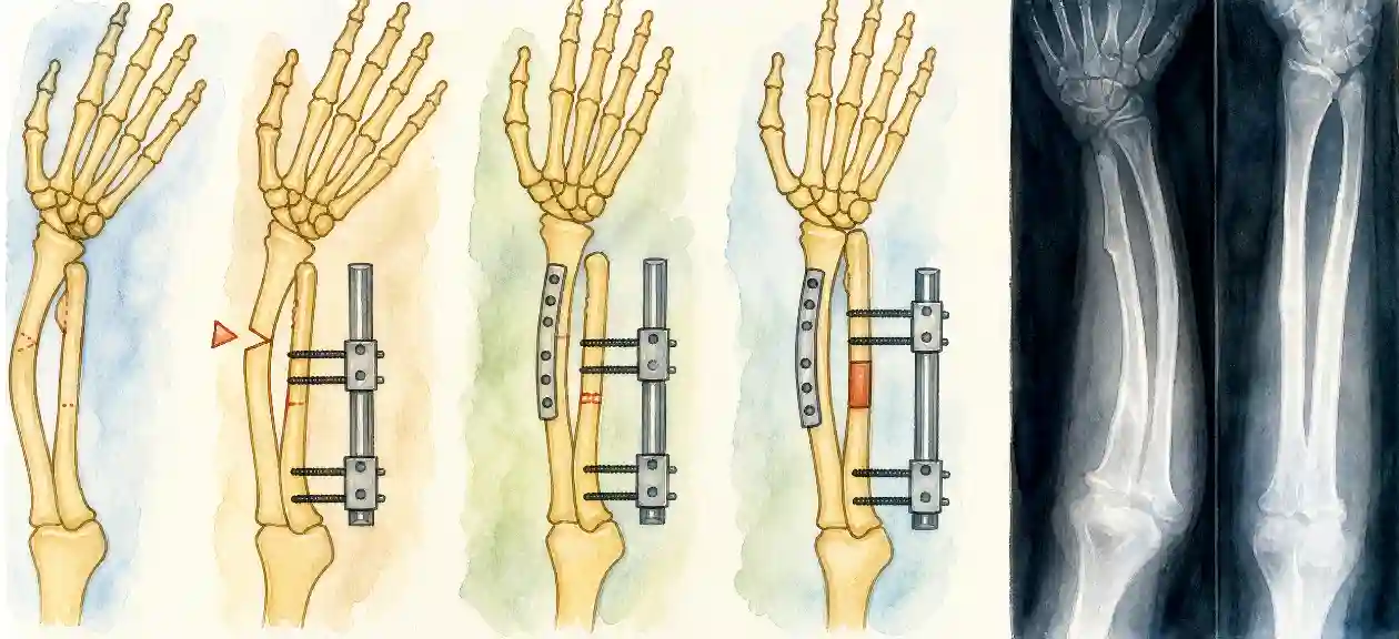
Ulna की lengthening, radius की osteotomy, या रुकावट डालने वाली गाँठ का निकालना — कलाई की shape और forearm के घुमाव (rotation) को सुधारने के लिए।
Limb lengthening / epiphysiodesis
लंबाई का फ़र्क़ ज़्यादा हो तो छोटी हड्डी को lengthen किया जाता है, या सही समय पर लंबी टाँग की growth plate रोक दी जाती है।
Not every lump has to come out. That is the most important line on this page. A lump that does not hurt and does not press on anything is measured and watched.
👀 Observation — the right answer for most children
A review every 6–12 months: the size of the lump, joint movement, limb lengths, and an X-ray when needed. Your child carries on playing normally. Once growth stops, the lump stops too.
✂️ When surgery is advised
- Pain that does not settle with rest, or a bursa that keeps coming back
- Pressure on a nerve or artery — numbness, weakness, an altered pulse
- Blocked joint movement — a knee or elbow that will not fully bend or straighten
- Increasing deformity — the forearm, ankle valgus, knock knee
- Any warning sign — growth after maturity, rapid enlargement, a thickening cap
- Daily interference — a lump that a shoe, a bag or sitting rubs constantly
🔧 The operations we use
Excision of the exostosis
The lump is removed at its base together with the entire cartilage cap and the perichondrium covering it. Take the whole cap and recurrence becomes uncommon.
Guided growth (8-plate)
A small plate on one side of the growth plate straightens the bone gradually, without cutting it. Excellent for ankle valgus and knock knee — but only while growth remains.
Forearm reconstruction
Lengthening the ulna, an osteotomy of the radius, or removing the offending lump — to restore the shape of the wrist and the rotation of the forearm.
Lengthening or epiphysiodesis
A significant length difference is treated by lengthening the short side, or by stopping the growth plate of the longer side at the right moment.
Operation के बाद — रिकवरी का रास्ता
After Surgery — The Recovery Road
यह एक सामान्य excision के बाद की तस्वीर है। Forearm reconstruction या lengthening में समय ज़्यादा लगता है, और उसकी planning अलग से की जाती है।
Operation का दिन
ज़्यादातर excision day-care या एक रात में हो जाते हैं। General anaesthesia, छोटा चीरा, गाँठ जड़ से निकाल दी जाती है।
घाव भरना
दर्द की दवा कुछ दिन। टाँके/dressing की जाँच। हल्का चलना-फिरना शुरू। पैर की surgery हो तो crutches कुछ दिन।
School और रोज़मर्रा
ज़्यादातर बच्चे 2 हफ़्ते के अंदर school लौट जाते हैं। Physiotherapy से joint का movement वापस आता है।
खेल-कूद वापस
हड्डी की जगह भर जाने के बाद धीरे-धीरे running और खेल। Contact sports डॉक्टर की सलाह पर।
Follow-up जारी
HME में नई गाँठें बन सकती हैं — इसलिए growth पूरी होने तक साल में एक बार जाँच जारी रहती है।
This is the picture after a straightforward excision. Forearm reconstruction and lengthening take longer, and are planned separately.
The day of surgery
Most excisions are day-care or a single night in hospital. General anaesthesia, a small incision, and the lump is taken from its base.
Wound healing
Pain relief for a few days, a wound check, and gentle movement from the start. Crutches for a few days if the leg was operated on.
School and daily life
Most children are back at school within two weeks. Physiotherapy restores full joint movement.
Back to sport
Running and games return gradually once the bone surface has healed. Contact sport on your surgeon's advice.
Follow-up continues
In HME new lumps can still appear, so a yearly review continues until growth is complete.
HME के साथ ज़िंदगी — माता-पिता के लिए
Living with HME — A Note for Parents
HME वाले बच्चे school जाते हैं, खेलते हैं, पढ़ते हैं, नौकरी करते हैं और सामान्य ज़िंदगी जीते हैं। ज़्यादातर को कुछ ही operations की ज़रूरत पड़ती है, और वे भी बरसों के अंतराल पर।
खेल-कूद
ज़्यादातर खेल खुले हैं। जहाँ गाँठ बाहर की तरफ़ है वहाँ padding या खेल बदलना काफ़ी है।
नियमित जाँच
Growth पूरी होने तक साल में एक बार; उसके बाद कोई नई तकलीफ़ हो तभी — पर warning signs हमेशा याद रखें।
Genetic counselling
शादी और परिवार की planning के समय बहुत मददगार — हर बच्चे को 50% chance का मतलब समझना ज़रूरी है।
बच्चे का मन
बड़े होते बच्चों को गाँठों को लेकर शर्म महसूस हो सकती है। खुलकर बात करना, और ज़रूरत पड़े तो counselling — इलाज का हिस्सा है।
Children with HME go to school, play, study, work and live ordinary lives. Most need only a handful of operations across childhood, often years apart.
Sport
Nearly everything stays open. Where a lump sits in an exposed spot, padding or a change of sport is usually enough.
Regular review
Yearly until growth is complete, then only if something new appears — but the warning signs are worth remembering for life.
Genetic counselling
Valuable when planning a family — the 50% chance for each child deserves to be properly understood, not guessed at.
How your child feels
Older children can feel self-conscious about visible lumps. Talking about it openly, and counselling when needed, is part of the treatment.
भ्रम बनाम सच्चाई
Myths vs Facts
"तेल की मालिश से गाँठ बैठ जाएगी।"
यह हड्डी है, गाँठ नहीं जो घुल जाए। मालिश से यह न घटती है न बढ़ती है — हाँ, रगड़ से दर्द ज़रूर बढ़ सकता है।
"हड्डी की हर गाँठ cancer होती है।"
Osteochondroma benign है। अकेली गाँठ में बदलाव 1% से भी कम मामलों में होता है — और X-ray/MRI से इसे पहचाना जा सकता है।
"जितनी जल्दी operation, उतना अच्छा।"
बिना तकलीफ़ वाली गाँठ में जल्दबाज़ी का फ़ायदा नहीं — बल्कि growth plate के पास जल्दी operation से नुक़सान का ख़तरा है। सही समय पर सही operation ज़्यादा अहम है।
"Operation के बाद गाँठ फिर से आ ही जाती है।"
पूरी cartilage cap निकल जाए तो दोबारा होना कम है। HME में कहीं और नई गाँठ बनना अलग बात है — वह recurrence नहीं है।
"Calcium की कमी से ये गाँठें बनती हैं।"
इसका खाने-पीने या calcium से कोई संबंध नहीं। यह growth plate की cartilage के खिसकने से बनती है, और HME में gene से।
"Massage with oil will make the lump go down."
It is bone, not a swelling that can dissolve. Massage neither shrinks nor grows it — though rubbing can certainly make it hurt more.
"Any lump on a bone means cancer."
An osteochondroma is benign. Change occurs in under 1% of solitary lumps — and imaging can identify it when it happens.
"The sooner it is operated, the better."
There is nothing to gain by rushing a painless lump, and operating close to an open growth plate carries its own risk. The right operation at the right time matters far more.
"It always grows back after surgery."
Recurrence is uncommon when the whole cartilage cap is removed. A new lump elsewhere in HME is a different thing altogether — not a recurrence.
"It is caused by a calcium deficiency."
Diet and calcium have nothing to do with it. It comes from displaced growth-plate cartilage, and in HME from an inherited gene.
Indore में जाँच और इलाज — क्या साथ लाएँ
Getting Checked in Indore — What to Bring
बच्चों की हड्डी की गाँठें growth plate के ठीक बगल में होती हैं — इसलिए इनका इलाज करने वाला डॉक्टर वही होना चाहिए जो बच्चों की growth को समझता हो। Dr. Gaurav Jain (MBBS, D.Ortho, DNB Orthopaedics, MNAMS; बच्चों की हड्डी रोग और स्पाइन सर्जरी में फेलोशिप) Indore में बच्चों की हड्डी रोग विशेषज्ञता के साथ exostosis और HME का इलाज करते हैं — excision, guided growth, forearm reconstruction और limb lengthening सहित।
- पुरानी सारी X-ray filmें — तारीख़ के हिसाब से (बदलाव देखने के लिए एक ही film से ज़्यादा ज़रूरी है पुरानी और नई की तुलना)
- MRI/CT की CD या films, अगर पहले हुई हों
- पहले हुए operation के discharge papers और biopsy report
- परिवार में किसी और को ऐसी गाँठें हों तो उनकी जानकारी — HME के लिए अहम
- बच्चे को किस काम में तकलीफ़ होती है, इसकी छोटी सी list — और हो सके तो एक video
Bone lumps in children sit right beside the growth plate — so they belong with a surgeon who thinks about growth every day. Dr. Gaurav Jain (MBBS, D.Ortho, DNB Orthopaedics, MNAMS; fellowship-trained in pediatric orthopedics and spine surgery) treats exostosis and hereditary multiple exostoses in Indore, including excision, guided growth, forearm reconstruction and limb lengthening.
- All previous X-ray films, in date order — comparing old with new tells us far more than any single film
- Any MRI or CT discs or films already done
- Discharge papers from earlier surgery, and any biopsy report
- Details of anyone else in the family with similar lumps — important for HME
- A short list of what your child struggles with, and a phone video if it helps show it
अक्सर पूछे जाने वाले सवाल
Frequently Asked Questions
Exostosis क्या है? / What is an exostosis?
हड्डी से निकली एक गाँठ जिसके ऊपर cartilage की cap होती है — medical नाम osteochondroma। यह growth plate के पास बनती है और joint से दूर की ओर बढ़ती है। बच्चों का सबसे common benign bone tumour। यह cancer नहीं है।
A bony outgrowth with a cartilage cap on top — properly called an osteochondroma. It forms beside the growth plate and points away from the joint. It is the commonest benign bone tumour of childhood, and it is not cancer.
क्या यह cancer है? / Is it cancer?
नहीं। यह benign है। बहुत कम मामलों में — अकेली गाँठ में 1% से कम, HME में लगभग 1–5% — बड़ी उम्र में cartilage cap में बदलाव (chondrosarcoma) हो सकता है। इसीलिए warning signs याद रखना ज़रूरी है, डरना नहीं।
No. It is benign. In a small minority — under 1% of solitary lumps and a reported 1–5% in HME — the cartilage cap can change in adult life into a chondrosarcoma. That is why the warning signs are worth knowing; constant worry is not needed.
गाँठ कब तक बढ़ेगी? / When does it stop growing?
जब तक बच्चा बढ़ रहा है। Puberty के बाद growth plates बंद होते ही गाँठ भी बढ़ना बंद कर देती है। अगर growth रुकने के बाद भी गाँठ बढ़े — तो तुरंत जाँच ज़रूरी है।
For as long as your child is growing. When the growth plates close after puberty, the lump stops too. A lump that grows after the skeleton has matured must be examined promptly.
HME क्या है और किसे होता है? / What is HME?
Hereditary Multiple Exostoses — कई हड्डियों पर कई गाँठें। EXT1 या EXT2 gene में बदलाव से होता है, autosomal dominant तरीक़े से। माता या पिता में से किसी एक को हो, तो हर बच्चे को 50% chance। ज़्यादातर बच्चों में 12 साल से पहले पता चल जाता है।
Hereditary Multiple Exostoses — many lumps across several bones, caused by a fault in the EXT1 or EXT2 gene and inherited in an autosomal dominant pattern. If one parent has it, each child has a 50% chance. Most children are diagnosed before the age of 12.
बाक़ी बच्चों की जाँच करवाएँ? / Should siblings be checked?
हाँ, अगर HME confirm हुआ है। भाई-बहनों और माता-पिता की simple clinical जाँच काफ़ी है — कई बड़ों को अपनी हल्की गाँठों का पता ही नहीं होता। Genetic counselling आगे की planning में मदद करती है।
Yes, once HME is confirmed. A simple clinical examination of siblings and parents is usually enough — many adults never realise they have mild lumps. Genetic counselling helps with planning ahead.
क्या हर गाँठ निकालनी पड़ेगी? / Does every lump need surgery?
नहीं। यह सबसे ज़रूरी बात है। जो गाँठ दर्द नहीं देती, किसी nerve पर दबाव नहीं डालती और movement नहीं रोकती — उसे सिर्फ़ नापा और देखा जाता है। Operation तब जब दर्द, दबाव, टेढ़ापन या warning sign हो।
No — and this matters most of all. A lump that does not hurt, does not press on anything and does not limit movement is simply measured and watched. Surgery is for pain, pressure, deformity or a warning sign.
कोई दवा है? / Is there a medicine for it?
ऐसी कोई दवा नहीं जो गाँठ को घटा दे या नई गाँठें रोक दे। दर्द के लिए सामान्य दर्दनिवारक, और रगड़ से बचाव — यही मदद करते हैं। Calcium या tonic का इससे कोई संबंध नहीं।
There is no medicine that shrinks a lump or prevents new ones. Simple pain relief and protecting the lump from friction are what help. Calcium supplements and tonics make no difference.
Operation कितना बड़ा होता है? / How big is the operation?
एक सामान्य excision आमतौर पर day-care या एक रात का काम है — general anaesthesia में छोटा चीरा, गाँठ जड़ से निकाल दी जाती है। ज़्यादातर बच्चे 2 हफ़्ते में school लौट जाते हैं। Forearm reconstruction या lengthening बड़ी surgery है, और उसकी planning अलग होती है।
A routine excision is usually day-care or one night — a small incision under general anaesthesia, and the lump comes off at its base. Most children are back at school within two weeks. Forearm reconstruction or lengthening is a bigger undertaking and is planned separately.
क्या गाँठ दोबारा आ सकती है? / Can it come back?
अगर पूरी cartilage cap और perichondrium निकल जाए तो दोबारा होना कम है। Cap का टुकड़ा छूट जाए या बच्चा बहुत छोटा हो तो खतरा थोड़ा बढ़ जाता है। HME में कहीं और नई गाँठ बनना recurrence नहीं है — वह बीमारी का स्वभाव है।
Recurrence is uncommon when the whole cartilage cap and its perichondrium are removed. Leaving a fragment of cap behind, or operating in a very young child, raises the risk a little. A new lump elsewhere in HME is not a recurrence — that is the condition doing what it does.
Surgery के risks? / What are the risks?
हर operation की तरह infection, खून बहना और anaesthesia के सामान्य risks। जगह के हिसाब से nerve या blood vessel के पास सावधानी ज़रूरी होती है, और growth plate के बहुत पास operation से growth प्रभावित हो सकती है — इसीलिए timing और सही specialist मायने रखते हैं।
The usual ones: infection, bleeding and the ordinary risks of anaesthesia. Depending on the site, a nerve or vessel may lie close by and needs care, and operating right next to a growth plate can affect growth — which is exactly why timing and the right specialist matter.
बच्चा खेल सकता है? / Can my child play sports?
ज़्यादातर मामलों में हाँ। बच्चे दौड़ते, खेलते और normal school life जीते हैं। खुली जगह पर बनी गाँठ को चोट लग सकती है या पतली डंडी वाली गाँठ टूट सकती है — वहाँ padding, खेल बदलना या उस एक गाँठ को निकालना काफ़ी होता है।
In most cases, yes. Children run, play and go through school normally. A lump in an exposed place can get knocked, and a thin stalked one can occasionally break — padding, a change of sport, or removing that one lump usually settles it.
Forearm छोटा क्यों रह जाता है? / Why is the forearm short and bent?
HME में ulna की growth जल्दी रुक जाती है जबकि radius बढ़ता रहता है — इससे forearm छोटा और मुड़ा हुआ हो जाता है और कलाई झुक जाती है। यह HME में operation का सबसे आम कारण है; इलाज में गाँठ निकालना, ulna lengthening या radius की osteotomy शामिल हो सकती है।
In HME the ulna often stops growing early while the radius carries on, so the forearm becomes short and bowed and the wrist tilts. It is the commonest reason for surgery in HME, and treatment may involve removing the lump, lengthening the ulna, or an osteotomy of the radius.
भविष्य कैसा रहेगा? / What does the future hold?
बहुत अच्छा। अकेली गाँठ वाले बच्चों को अक्सर एक बार के इलाज के बाद कुछ नहीं चाहिए। HME वाले बच्चे भी पढ़ाई, नौकरी और सामान्य ज़िंदगी जीते हैं — बस growth के दौरान नज़र रखनी होती है और ज़रूरत पड़ने पर कुछ operations।
Good. Children with a solitary lump usually need nothing further once it is dealt with. Children with HME study, work and live ordinary lives too — they need watching through the growing years and, when necessary, a few well-timed operations.
किस डॉक्टर को दिखाएँ? / Which doctor should we see?
बच्चों की हड्डी रोग विशेषज्ञ (pediatric orthopedic surgeon) — क्योंकि ये गाँठें growth plate से जुड़ी होती हैं और इलाज बच्चे की growth के हिसाब से तय होता है। Indore में Dr. Gaurav Jain (MBBS, D.Ortho, DNB Orthopaedics, MNAMS) exostosis और HME दोनों का इलाज करते हैं।
A pediatric orthopedic surgeon — these lumps are tied to the growth plate, and the plan depends on how much growing is left. In Indore, Dr. Gaurav Jain (MBBS, D.Ortho, DNB Orthopaedics, MNAMS) treats both exostosis and hereditary multiple exostoses.
📞 गाँठ दिखी है? Dr. Gaurav Jain से जाँच करवाएँ - Indore!
📞 Found a lump? Get it checked by Dr. Gaurav Jain - Indore!
Indore के Pediatric Orthopedic Surgeon | Exostosis, HME, Deformity Correction व Limb Lengthening Pediatric Orthopedic Surgeon in Indore | Exostosis, HME, Deformity Correction & Limb Lengthening
📞 अभी Call करें 📞 Call Nowपुरानी X-ray साथ लाएँ — पुरानी और नई film की तुलना ही सबसे ज़्यादा बताती है। Bring the old X-rays — comparing them with today's film tells us the most.